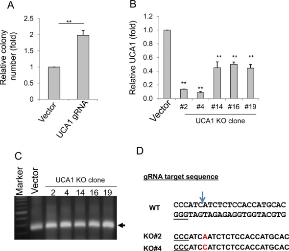
Figure 4.

Generation of UCA1 knockout in HCT-116 cells. (A) Colony formation for vector and UCA1 KO for a mixed pool after puromycin selection. (B) Detection of UCA1 expression in UCA1 KO clones by qRT-PCR. (C) Detection of UCA1 target region in all five clones using genomic DNA as a template and primers UCA1-inside-5.3 and UCA1-inside-3.3. (D) Detection of a single nucleotide insertion (in red) in UCA1 KO#2 and #4, by DNA sequencing. The arrow indicates the cleavage site by Cas9. Underlined GGG is PAM sequence. Values in (A) and (D) are means of ± SE (n = 3). **P < 0.01.
