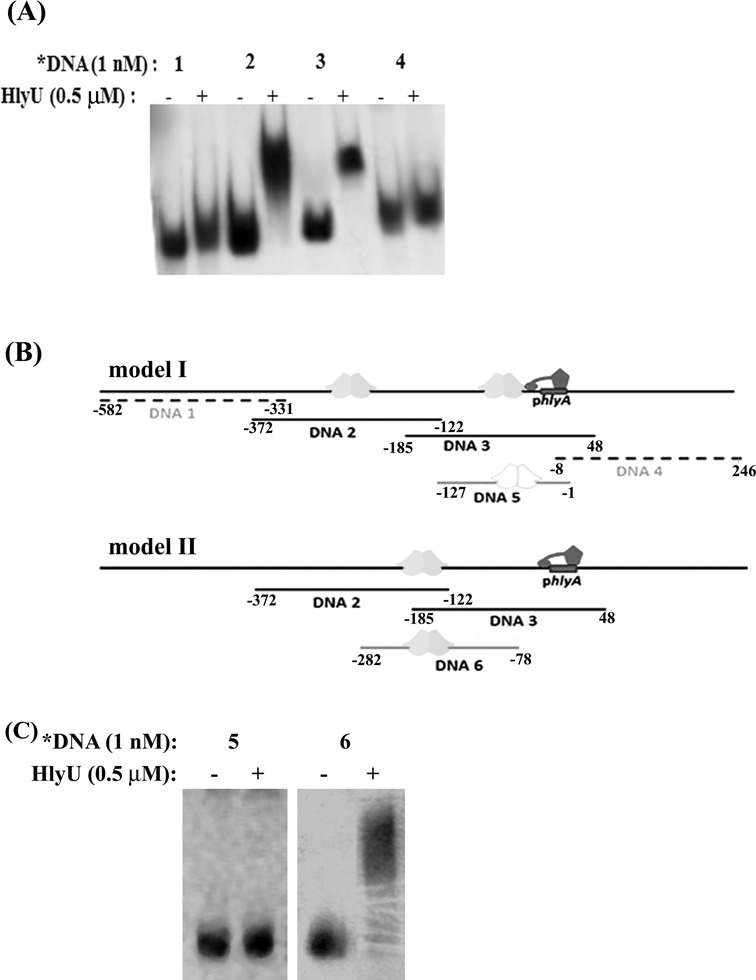
Figure 1.

The binding of HlyU_Vc to the upstream region of the hlyA gene. (A) EMSA showing protein binding to DNA sequences 1–4 (Table 1). (B) Models for HlyU_Vc binding that can account for shift in mobility of both DNA 2 and DNA 3. (model I) There are separate binding sites for HlyU_Vc on both DNA 2 and DNA 3, in which case HlyU_Vc should show shift with DNA 5. (model II) HlyU_Vc binds to the overlapping regions of DNA 2 and DNA 3, in which case HlyU_Vc should show shift with DNA 6. The dimeric HlyU_Vc, hlyA promoter (phlyA) and the RNA pol are represented as cartoons. (C) EMSA showing protein binding to DNA sequences 5–6.
