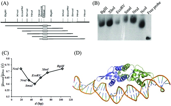
Figure 6.

(A) Restriction map of the probes used in the DNA bending assay (from vector pBend4); the oblong indicates location of the DNA 6 in the DNA probe. (B) Circular permutation assay to determine deviation from linearity of DNA on binding to HlyU_Vc. (C) Plot of μbound/μfree (‘r’, the ratio of mobility (μ) of the bound form and the free probe for each restriction enzyme digested fragment with the DNA6 insert) versus the position (d, in bp) of the DNA insert from the 5′ end of each restriction fragments. The minimum and maximum values of the ratio were used to calculate α (bend angle) according to Equation (1). (D) DNA-HlyU_Vc model after 10 ns MD simulation.
