
Figure 2.
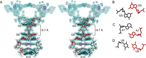
Parallel-stranded duplex. (A) Stereoview of residues 1–10 from two monomers that form a ps duplex shown with 2mFo–DFc electron density contoured at 0.75 σ. Parallel-stranded duplex residues are labeled. Residues outside the duplex region (A1–U3) are semi-transparent. Anomalous difference electron density contoured at 5 σ (violet surface) corresponds to bromine atoms used for phasing. Water molecules are shown as red spheres. The gap between residues G5 and G6 is 6.7 Å. (B–D) Individual base pairs showing hydrogen bonding between identical residues. (B) N2–N3 sugar edge interactions between G5–G5, G6–G6 and G9–G9 homo base pairs. (C) N6–N7 Hoogsteen interactions between A7–A7 and A10–A10. (D) Symmetric N3–O4 hydrogen bonding observed for U8–U8 homo base pair.