
Figure 4.
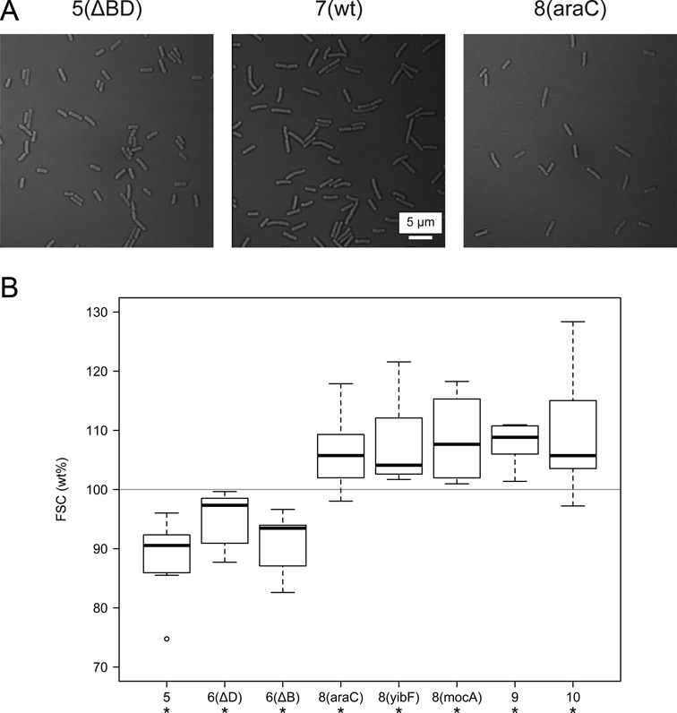
(A) Microscopic images of selected strains. (B) The size (forward scatter, FSC) of the modified cells was measured by flow cytometry and compared to the wt in each measurement. Center lines show the medians of relative size; box limits indicate the 25th and 75th percentiles; whiskers extend 1.5 times the interquartile range; and outliers are represented by circles. Results are based on seven independent experiments (20 000 cells per experiment). Asterisks denote significant difference compared to the wt (*P < 0.05, paired samples t-test).