Fig. 2.

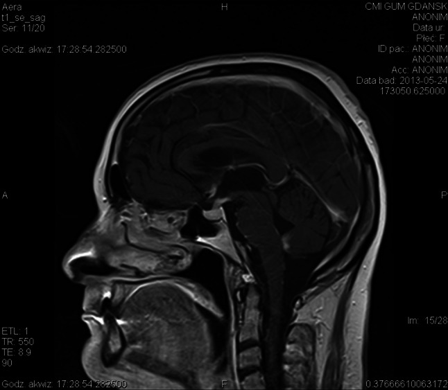
Neurosarcoidosis in MRI brain in sagital plane: T1-weighted contrast-enhanced MR image shows basal leptomeningeal enhancement and an extensive enhancement of the pituitary gland and stalk, which is markedly enlarged

Neurosarcoidosis in MRI brain in sagital plane: T1-weighted contrast-enhanced MR image shows basal leptomeningeal enhancement and an extensive enhancement of the pituitary gland and stalk, which is markedly enlarged