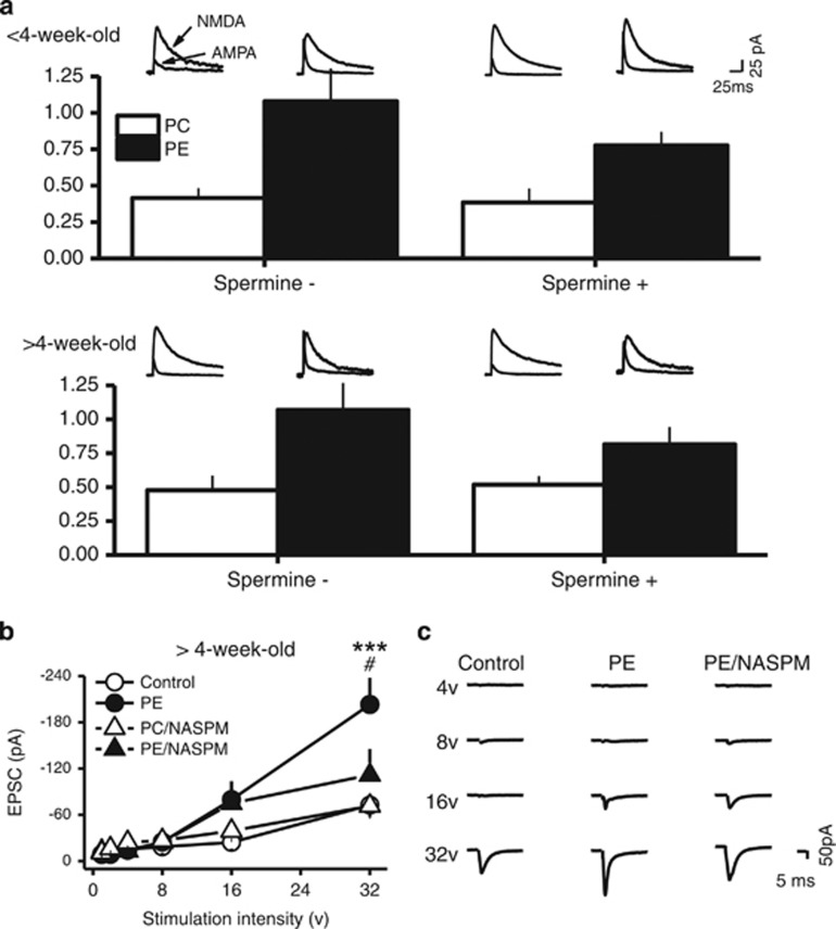
Figure 5.
Prenatal ethanol exposure increases the strength of excitatory synapses of VTA DA neurons. (a) PE enhanced AMPAR/NMDAR ratio in animals <4-week-old and >4-week-old. Bar graph depicting the average AMPAR/NMDAR ratios obtained in control and PE animals. Note an increase in AMPAR/NMDAR ratio in PE animals in both age groups and this effect was not altered by the absence or presence of spermine in the internal solution, suggesting endogenous polyamines in VTA DA neurons. Superimposed AMPAR- and NMDAR-EPSCs traces recorded at +50 mV in control (left traces) and PE (right traces) animals are presented above the bar graphs. (b) Input-output curves of AMPR-EPSCs obtained in control and PE-exposed animals >4-week-old. Prenatal ethanol exposure significantly shifted the input-output I-O curve to the left. Specifically, PE led to increased AMPAR-EPSC amplitude when stimulation intensity reached 32 V. This effect was no longer observed in the presence of NASPM. (c) Sample traces of evoked AMPAR-EPSCs at different stimulation intensities in control and PE animals and in the presence of NASPM in PE animals. *P<0.05; **P<0.01, differences between control and PE groups. #P<0.05, differences between vehicle and NASPM in PE animals.

