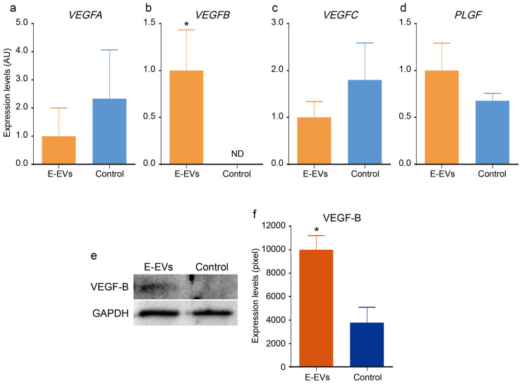
Figure 6. Effects of E-EVs in cerebrovascular pericytes.
(a–d) The mRNA levels of the VEGF family members, VEGFA, VEGFC, and PLGF in the E-EV-supplemented and non-stimulated control groups did not differ significantly (a, c, and d). In contrast, VEGFB was significantly upregulated in the E-EV-supplemented group compared to the non-stimulated control group (b) (n = 3, P < 0.05). (e and f) VEGF-B protein levels were measured by western blotting (e), using GAPDH as an internal control. VEGF-B protein was significantly upregulated in the E-EV-supplemented group compared to the non-stimulated control group (f) (n = 3, P < 0.05).

