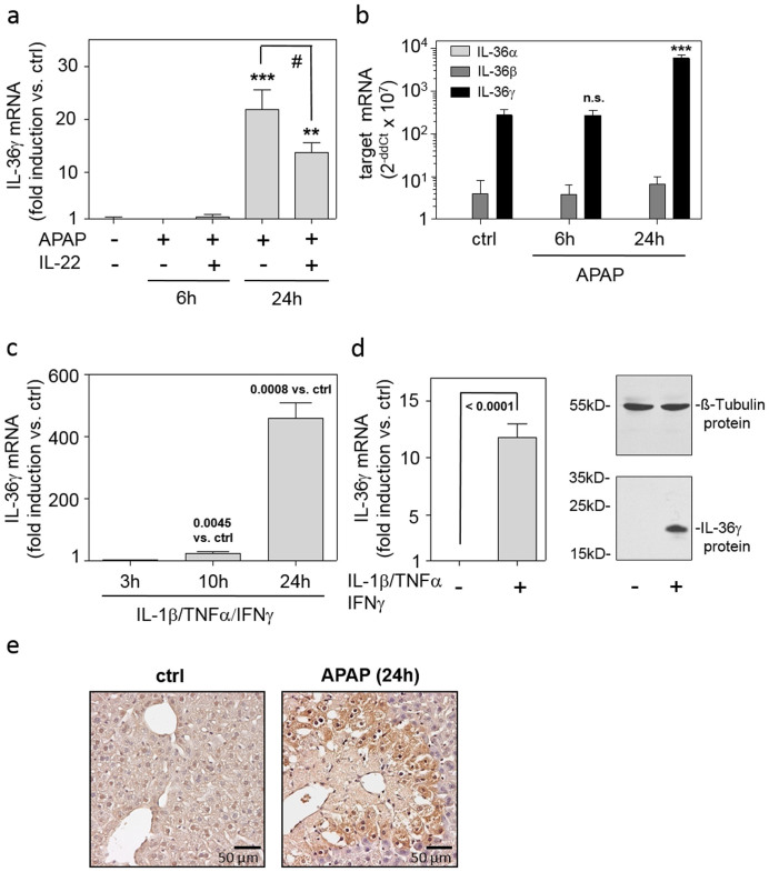
Figure 1. Expression of IL-36γ in murine APAP-induced liver injury and inflamed hepatocytes.
(ab) Mice received PBS (n = 6) or APAP or (where indicated) APAP/IL-22 (6 h (n = 6), 24 h (n = 9)). (a) Hepatic IL-36γ mRNA was determined by realtime PCR. Target mRNA was normalized to that of GAPDH (means ± SEM versus ctrl; **p < 0.01, ***p < 0.001 versus ctrl; #p < 0.05). (b) Hepatic IL-36αβγ mRNAs were determined by realtime PCR. Target mRNA was normalized to that of GAPDH and is shown as raw data (2−ddCt × 107; means ± SEM; ***p < 0.001 versus ctrl of the same target gene; n.s., not significant). (ab) Statistical analysis, one-way analysis of variance with post hoc Bonferroni correction. (c) Murine primary hepatocytes were kept as unstimulated control (ctrl) or stimulated with IL-1β/TNFα/IFNγ (each 50 ng/ml). IL-36γ mRNA expression was determined by realtime PCR. Target mRNA was normalized to that of GAPDH (means ± SEM versus ctrl; n = 3). (d) Huh7 cells were kept as unstimulated ctrl or stimulated with IL-1β/TNFα/IFNγ (each 50 ng/ml). After 3 h (left panel) or 24 h (right panel), IL-36γ mRNA (left panel) or protein (right panel) was determined by realtime PCR or immunoblotting, respectively. Target mRNA was normalized to that of GAPDH (means ± SD versus ctrl; n = 8). One representative immunoblot of five independently performed experiments is shown. (cd) Statistical analysis, Student's t-test versus ctrl at the respective time point. (e) Representative murine liver immunohistochemistry of IL-1Rrp2 at 24 h after APAP application (left panel: ctrl; right panel: APAP).

