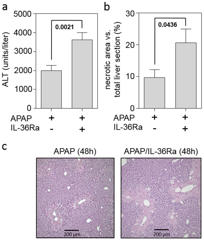
Figure 4. Application of IL-36Ra exacerbates tissue damage in late murine APAP-induced liver injury.
(a) Mice received either APAP (n = 16) or APAP/IL-36Ra (n = 16) and were maintained for 48 h. Thereafter, serum ALT activity was determined and is depicted as units/liter (means ± SEM). (b) Mice received either APAP (n = 8) or APAP/IL-36Ra (n = 8). Statistical analysis of necrotic areas in H&E-stained liver sections after 48 h. (ab) Statistical analysis, Student's t-test. (c) Representative liver sections (H&E staining) 48 h after the onset of APAP intoxication.

