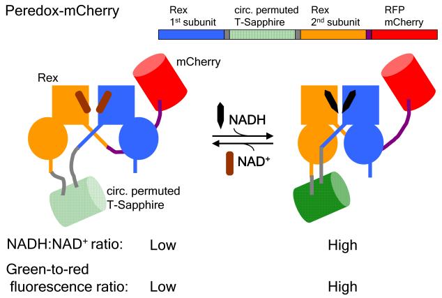
Fig. 1.
Schematic showing the design of the fluorescent sensor of the cytosolic NADH-NAD+ redox state, Peredox-mCherry: A circularly permuted GFP T-Sapphire (green) is interposed between the two Rex subunits (blue and orange), with a C-terminal RFP mCherry to normalize for the green fluorescence. Upon binding to NADH (black) but not NAD+ (brown), Rex undergoes a conformational change that is coupled to an increase in the GFP fluorescence. Thus, the green-to-red fluorescence ratio increases with NADH:NAD+ ratio. Adapted from Hung et al., 2011.

