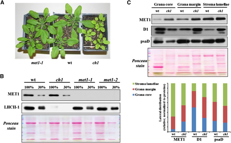
Figure 11.
Accumulation of MET1 and LHCII in ch1 and met1 Alleles and the Wild Type.
(A) Plants used for immunoblotting. Bar = 3 cm.
(B) Immunoblot of MET1 and LHCII-1 of thylakoids isolated from ch1, met1-1, met1-2, and the wild type. The gel was loaded based on equal total leaf protein (100% = 10 μg).
(C) Distribution of MET1 across the different thylakoid membrane regions in the wild type and the ch1 mutant. Thylakoid proteins were solubilized with digitonin, and grana, grana margins, and stroma lamellae were fractionated by ultracentrifugation and analyzed by SDS-PAGE (15 μg protein per lane). Immunoblots for MET1, D1 (PSII core), and PsaD (PSI core) protein and the Ponceau stain are shown. The bar diagram shows the relative distribution of MET1, D1, and PsaD across the thylakoid regions in the wild type and ch1 with signals normalized to the total signal for each protein within each genotype.

