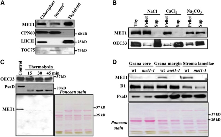
Figure 4.
MET1 Is a Stroma-Exposed Peripheral Thylakoid Protein Enriched in Stroma Lamellae.
(A) Localization of MET1 within the chloroplasts. Total chloroplast, thylakoid membranes, and stroma were used for immunoblotting with MET1 antiserum. Antisera against stromal CPN60, thylakoid LHCII, and outer envelope protein TOC75 were used as markers. Asterisk indicates that the stroma still contains envelopes, as it was collected as the supernatant from broken chloroplasts following a 10-min spin at ∼18,000g.
(B) Salt washing of thylakoid membranes. The membrane was sonicated in the presence of NaCl, CaCl2, and Na2CO3 and incubated on ice for 30 min before centrifugation to separate soluble and membrane fractions. Ten micrograms of proteins from supernatant and pellets was loaded on the SDS-PAGE gel. For control, thylakoids without any treatment of salt or sonication were used.
(C) Thermolysin treatment of thylakoid membranes. Thylakoids isolated from wild-type chloroplasts were treated with thermolysin for 15, 30, and 45 min on ice and then immunoblotted. Proteins were separated by SDS-PAGE and immunoblotted with OEC33 (PsbO), PsaD, and MET1 antisera. The Ponceau-stained image prior to blotting is shown. Ten micrograms of protein was loaded in each lane.
(D) Distribution of MET1 across the different thylakoid membrane regions. Thylakoid proteins were solubilized with digitonin and grana, grana margins, and stroma lamellae were fractionated by ultracentrifugation and analyzed by SDS-PAGE. Ten micrograms of protein was loaded in each lane. Immunoblots for MET1, D1, and PsaD protein are shown. Chlorophyll a/b ratios with standard deviations in parentheses for the wild-type fractions were: 3.4 (±0.41), 2.5 (±0.01), 3.0 (±0.15), and 6.6 (±0.15) for thylakoids, grana core, grana margins, stromal lamellae, respectively. The chlorophyll a/b ratios for the met1 fractions are: 3.6 (±0.5), 2.5 (±0.07), 3.7 (±0.34), and 5.6 (±0.94) for thylakoids, grana core, grana margins, and stromal lamellae, respectively. Ponceau-stained blots are shown for (C) and (D).

