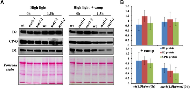
Figure 9.
Degradation of PSII Core Proteins after High Light Treatment.
Detached leaves of the wild type, met1-1, and met1-2 grown at 100 μmol photons m−2 s−1 (16 h light/8 h dark) exposed for 1.5 h to high light (1200 μmol photons m−2 s−1) in the absence or presence of the translational inhibitor chloramphenicol. Proteins were extracted from detached leaves and separated by SDS-PAGE gel and subjected to immunoblot with antisera against the PSII core proteins D1, D2, and CP43. Equal amounts of protein (10 μg) were loaded in each lane. (A) shows representative immunoblots and Ponceau stains, whereas (B) shows the average ratios of D1, D2, and CP43 proteins for the wild type and met1 before and after 1.5 h light stress. Data for the met1-1 and met1-2 alleles were averaged within each replicate. Standard deviations are indicated (n = 3). Two-tailed Student’s t tests showed significant differences at P < 0.05 for CP43 and at P < 0.1 for D2 in met1 with chloramphenicol.

