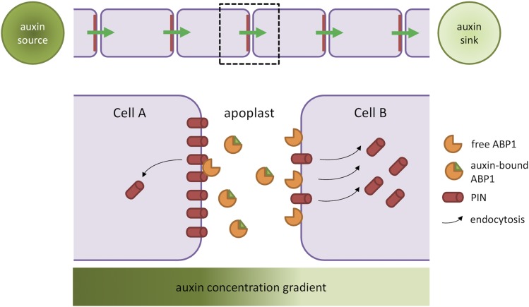
Figure 3.
A Model of Auxin Transport Canalization by Extracellular Auxin Perception by ABP1.
This figure shows an update of the model presented by Wabnik et al. (2010). PIN proteins gradually polarize to form a canal of auxin flow connecting auxin source to the sink. Two neighboring cells share an apoplastic pool of ABP1 molecules. ABP1 exists in auxin-free and auxin-bound states, whereby it promotes endocytosis or is inactive, respectively. Due to an auxin concentration gradient across the apoplastic space, cell A (closer to the auxin source) experiences higher apoplastic auxin levels and fewer auxin-free ABP1 and thus has low PIN endocytosis rates, resulting in stabilization of PIN at the plasma membrane. The extracellular space near cell B has lower auxin concentration and more free ABP1 molecules that promote PIN removal from the plasma membrane.

