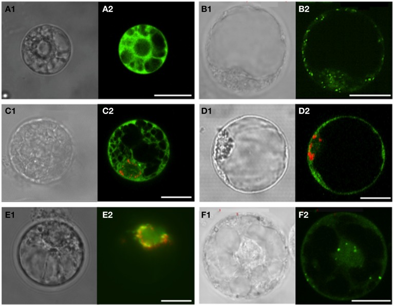
Figure 2.
Sub-cellular localization of GFP fused to the C-terminal sequences of Vitis vinifera cv Touriga Nacional SERAT proteins. Electroporation was carried out in V. vinifera protoplasts isolated from cell cultures. The signal expression and localization of GFP and RFP was observed between 24 and 48 h incubation by confocal laser scanning or epifluorescence microscopy. (A) Plasmid pFF19-GFP used as control for localization into the cytosol; (B) SHMT-GFP carrying the transit peptide of Serine hydroxymethyltransferase (SHMT) from Arabidopsis as a control for mitochondria targeting; (C,D) VvSERAT1;1-GFP and VvSERAT3;1-GFP co-transformed with VsRSS:RFP carrying the transit peptide of pea Rubisco small subunit as a control for plastidic localization, respectively. Both VvSERAT isoforms show cytosolic localization; (E) VvSERAT2;1-GFP and VsRSS:RFP co-localizing in the plastids; (F) VvSERAT2;2-GFP localized In the mitochondria and the cytosol. Letters followed by 1, Protoplasts observed in contrast phase microscopy; Letters followed by 2, Confocal laser scanning except for E2 observed by epifluorescence microscopy. Scale bars = 20 μm.

