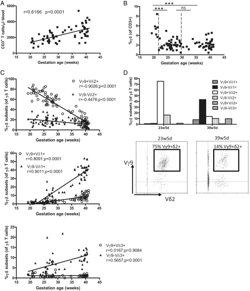
Fig. 1.
Human fetal peripheral blood is highly enriched for the presence of Vγ9Vδ2 T cells around 20 wk gestation. (A) Absolute numbers of T cells per microliter of blood as determined by flow cytometry on fresh blood using Trucount tubes (n = 64); the linear regression line is shown together with its r and P values. (B) Percentage of γδ T cells (of CD3+ lymphocytes) according to gestational age; total n = 87; comparisons are shown between 19w2d–21w2d (n = 7), 21w5d–29w5d (n = 40), and 30w3d–41w1d (n = 40). ***P < 0.001; ns, not significant. (C) Percentages of γδ T-cell subsets of γδ+CD3+ lymphocytes according to gestational age; linear regression lines are shown with their corresponding r and P values. (D) Flow cytometry data on γδ subset percentages of γδ+CD3+ lymphocytes for one fetus from which we obtained blood both at 23w5d and at 39w5d gestation; these data are representative fo the data for three different fetuses from which blood samples were derived at two different gestational times; in the lower panel, the gate is put on γδ+CD3+ lymphocytes.

