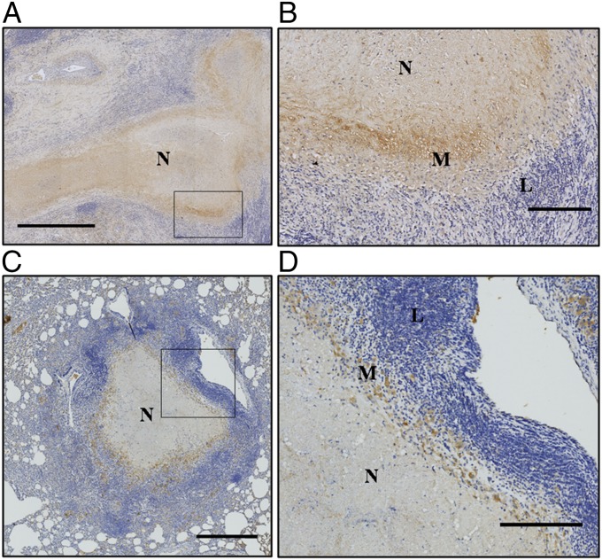
Fig. 1.
Human and rabbit granulomas express VEGF. Representative line-scanned histological sections from human patients after lung tissue resection surgery (A and B) and from necropsied rabbits (C and D) show VEGF expression (brown) in the cellular layers surrounding the central necrosis of necrotizing (N) granulomas in humans (A) and rabbits (C), with magnified regions shown in B and D. Macrophages (M) express higher levels of VEGF than lymphocytes (L) in granulomas. (Scale bars: A, 1 mm; B and D, 200 μm; C, 500 μm.)

