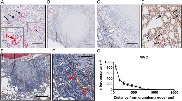
Fig. 2.
TB granuloma blood vessels are morphologically abnormal and have heterogeneous spatial densities. Human resection tissues (A–C) and rabbit tissues from an experimental MTB infection (D–F) labeled with CD31 (brown, open red triangles) and αSMA (pink, closed red triangles) are shown. The MVD is higher in human (A and Inset) and rabbit (D) normal lung tissue adjacent to lesions than in human (B and C) and rabbit (E and F) granulomas. Staining for endothelial cells revealed that in both human (B and C) and rabbit (E and F) granulomas, blood vessels are restricted to the granuloma periphery and tend to be absent from the central region, as demonstrated by quantification of MVD (the number of CD31-positive microvessels per square millimeter of viable tissue) in rabbit granulomas (G). Granuloma vessels are often compressed along the lesion periphery and collapsed within the interior (C and F, white arrows), as indicated by a positive CD31 stain and lack of a visible open vessel lumen, unlike the open, often αSMA-covered vessels in the normal lung (A and D, black arrows). αSMA costaining with CD31 shows that granuloma-associated blood vessels lack significant pericyte coverage in both human and rabbit tissues. (Scale bars: A, C, D, and E, 200 μm; A, Inset, 20 μm; B, 500 μm; F, 50 μm.)

