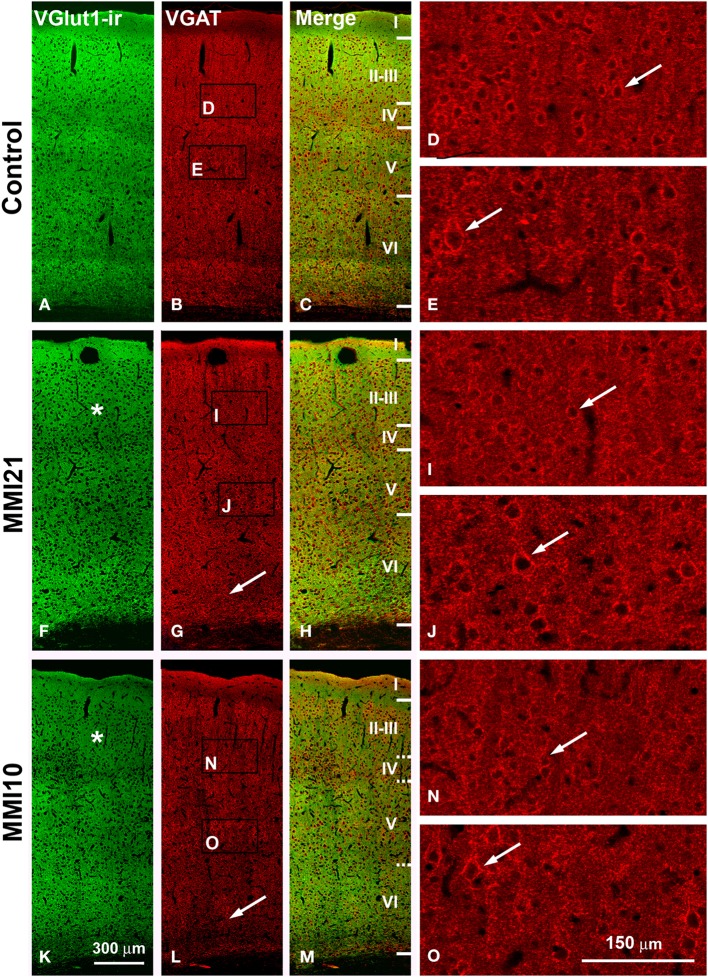
Figure 13.
Confocal immunolabeling in the somatosensory cortex of C and MMI pups. Confocal photomicrographs showing VGluT1-ir (green labeling; A,F,K), VGAT-ir (red labeling; B,G,L) and merged images (C,H,M) in the somatosensory cortex of C (A–E), MMI21 (F–J) and MMI10 (K–O) pups at P50. Asterisks (*) point to supragranular VGluT1-ir labeling in MMI (F,K) pups. Perisomatic VGAT-ir boutons are indicated by arrows. Boxes in (B,G,L) show the location of the corresponding enlarged figures. Note the decreased density and smaller size of perisomatic VGAT-ir boutons (arrows) in layers II-III and V in MMI10 pups (compare N,O with D,E,I,J). Same scale for (A–C, F–G, K–M) and for (D,E,I,J,N,O).

