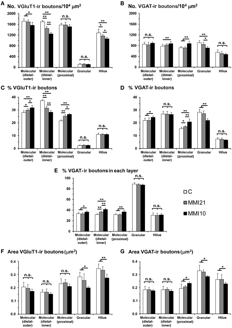
Figure 8.
VGluT1-ir and VGAT-ir bouton distribution in DG of C and MMI pups. Histograms showing the VGluT1-ir and VGAT-ir bouton distribution in DG of C and MMI pups. Note the deceased VGlut1-ir bouton density and percentage in the distal-inner molecular layer of MMI compared to C pups (A,C). The VGAT-ir bouton density and percentage increased in the proximal molecular layer and decreased in the granular layer of MMI10 pups, and increased in the proximal molecular layer of MMI21 pups (B,D). The VGAT-ir bouton percentage in each layer increased in the MMI10 distal (outer and inner) and proximal molecular and granular layers, and in the MMI21 distal-inner molecular layer (E). The VGluT1-ir and VGAT-ir bouton area was smaller in the MMI10 granular layer and hilus (F,G). n.s. indicates not significant differences; (*) and (**) indicate significant differences, P ≤ 0.05 and P ≤ 0.001, respectively.

