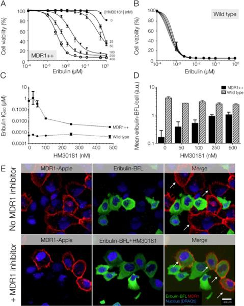
Fig. 5. Reversal of MDR1-mediated drug resistance in vitro using the experimental MDR1 inhibitor, HM30181.
(A) A third-generation MDR1 inhibitor, HM30181 (16), restored sensitivity to eribulin in a dose-dependent manner in vitro in HT1080 cells over-expressing MDR1. (B) HM30181 does not alter the cytotoxicity of eribulin in the drug-sensitive parent cell line, HT1080 (human fibrosarcoma). (C) Sensitivity of resistant and wild-type HT1080 cells to eribulin in the presence of HM30181. Data in (A to C) are reported as averages of triplicate experiments. (D) Cellular eribulin-BFL uptake in resistant (MDR1++) and wild-type cells in response to increasing concentrations of HM30181. Single-cell levels of fluorescence drug were determined by confocal microscopy, where all images were analyzed at same exposure settings. Per-cell count was determined using the DRAQ5 nuclear label. Data are means ± SEM (an average of n = 38 cells evaluated per condition). (E) Eribulin-BFL is not taken up by MDR1-expressing cells (white arrows) in a mixed population of HT1080 cells. However, incubating resistant cells with HM30181 allowed eribulin influx (white arrows). (For the effects of other MDR1 inhibitors, see fig. S10.) Images were acquired at identical exposure settings. The nuclear stain is DRAQ5. Images are representative of n = 5 FOV.

