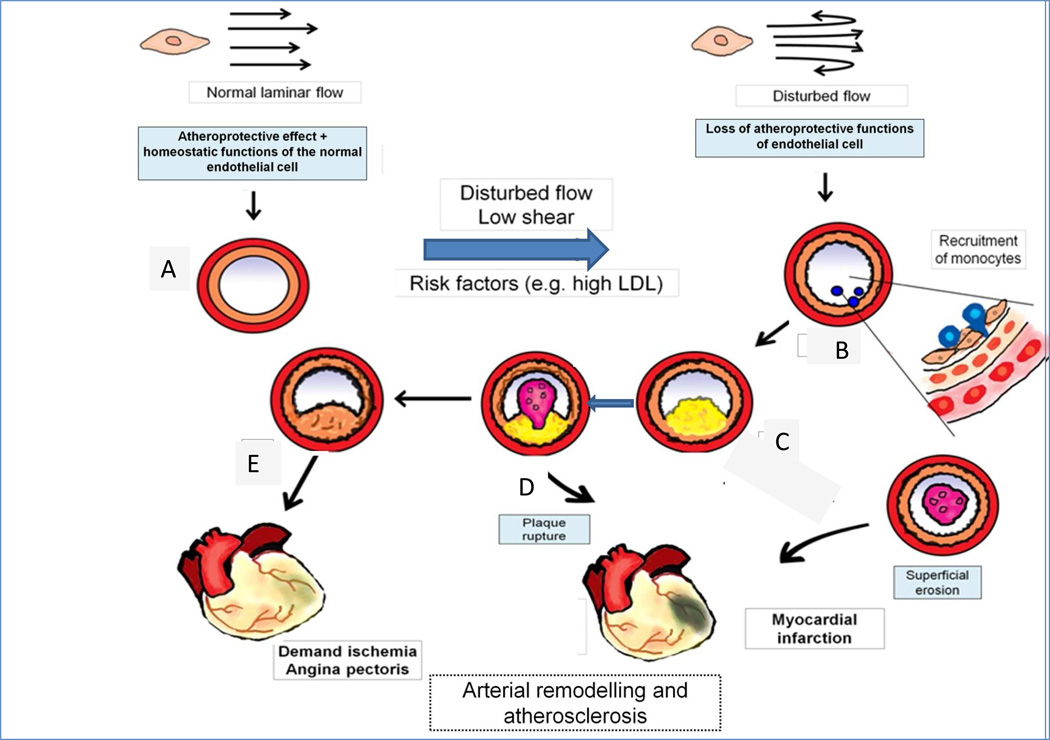
Figure 1. Positive and negative arterial remodelling influences the clinical consequences of atherosclerosis.
Normal laminar shear stress (upper left) elicits atheroprotective and homeostatic functions of endothelial cells (ECs). These functions maintain normal arterial caliber and properties (A). Disturbed blood flow in endocardial disease is shown by the reversed arrows in the upper right. In (B) the red circle portrays the tunica media, and the orange circle indicates the intima of the artery. Disturbed flow promotes the recruitment of monocytes, as depicted in the nascent plaque (B), where monocyte diapedesis (in blue) penetrates between ECs to form a thin-capped, lipid-rich inflamed plaque (C), which can rupture and cause a thrombus (D), leading to myocardial infarction, indicated by cyanosis in the heart diagram. Alternatively the plaque in E can undergo constrictive remodelling to promote flow-limiting stenosis (E) that can cause demand ischaemia and angina pectoris

