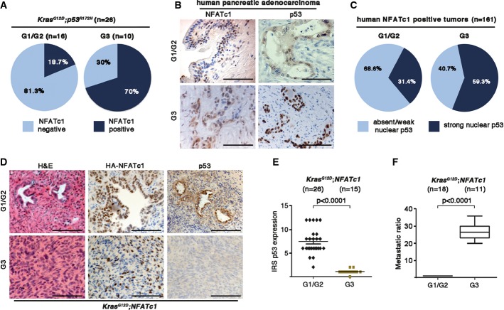
Figure 1.
- Statistical analysis of NFATc1 expression with respect to tumor grading in KPC mice tissues.
- Immunohistochemical detection of NFATc1 and p53, illustrating G1-G2 and G3 in human PDAC tissues. Scale bars, 100 μm.
- Statistical illustration of TMA analysis demonstrating NFATc1 strong nuclear positive tumors (n = 161 patients) with respect to functional status of p53 expression in human PDAC tissues.
- Representative H&E-stained sections as well as immunohistochemical detection of HA-NFATc1 and p53 in well- and poorly differentiated pancreatic tissues of KNC mice. Scale bars, 100 μm.
- Quantification of p53 expression with respect to the tumor grading by evaluating both the intensity of immunostaining and the number of cells with overexpression by means of an immunoreactivity score (IRS) in KNC mice (every dot represents one mouse).
- Incidence of liver metastases in KNC mice (mean values ± SD).

