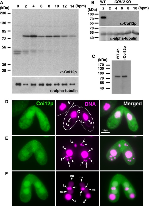
Figure 2.

- Proteins extracted from wild-type cells at the indicated time point of conjugation (hpm) were analyzed by Western blot with the newly established anti-Coi12p antibody (top) and with an anti-alpha-tubulin antibody (bottom). The positions of the protein molecular weight markers are indicated at the left.
- Proteins extracted from wild-type cells at 2 hpm and from the COI12KO cells at the indicated time of conjugation (hpm) were analyzed by Western blot as described in (A).
- Proteins extracted from wild-type cells at 4 hpm and recombinantly expressed Coi12p from E. coli were analyzed by Western blot as described in (A).
- Immunofluorescence staining of wild-type cells using the anti-Coi12p antibody. Coi12p localizes to the cytoplasm and the MAC in early stage (D, meiotic prophase), mid-stage (E, nuclear exchange), and late stage (F, nuclear alignment) of conjugation. The MICs (i), parental MACs (a) and newly developed MACs (na) are marked with arrowheads. Vegetative (V) and conjugating (C) cells are circled with dotted lines in (D).
