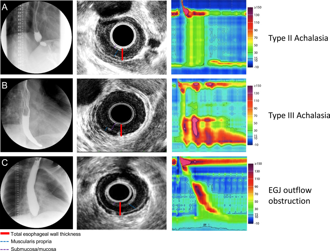
Figure 1. Representative EUS images in patients with achalasia.
Row (A) Fluoroscopy, EUS and HRM are shown in a patient with typical type II achalasia. The fluoroscopy revealed retained barium with narrowing toward the EGJ. Endosonographically, the esophageal wall was thickened with contribution from a hypoechoic muscular layer and isoechoic submucosal layer. Row (B). Type III achalasia. Note the typical early latency contraction shown on HRM. Retained contrast was noted on fluoroscopy. EUS revealed a thickened esophageal wall with the majority comprised of an isoechoic mucosal/submucosal layer. Row (C). A patient with EGJ outflow obstruction. High resolution manometry revealed intact peristalsis with elevated IRP. Fluoroscopy revealed a high column of retained barium. EUS revealed a predominately thickened hypoechoic muscular layer.

