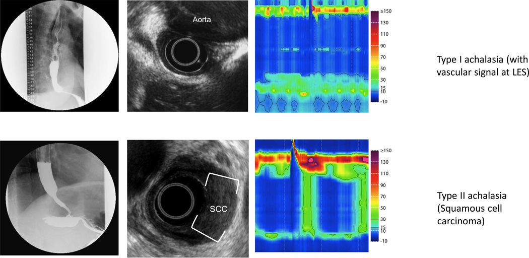
Figure 2. Pseudoachalasia identified on EUS.
Row (A) Fluoroscopy reveals a typical achalasia like pattern. High resolution manometry confirmed absent peristalsis with elevated IRP, however, careful inspection revealed a vascular artifact. EUS confirmed compression of the EGJ by the aorta with loss of the typical plane between the aorta and esophagus. CT confirmed a massively dilated thoracic aneurysm. Row (B) Fluoroscopy and manometry reveal achalasia (type II pattern). This patient had normal standard endoscopy. EUS revealed asymmetric thickening of the distal esophagus with an isoechoic lesion. FNA confirmed squamous cell carcinoma.

