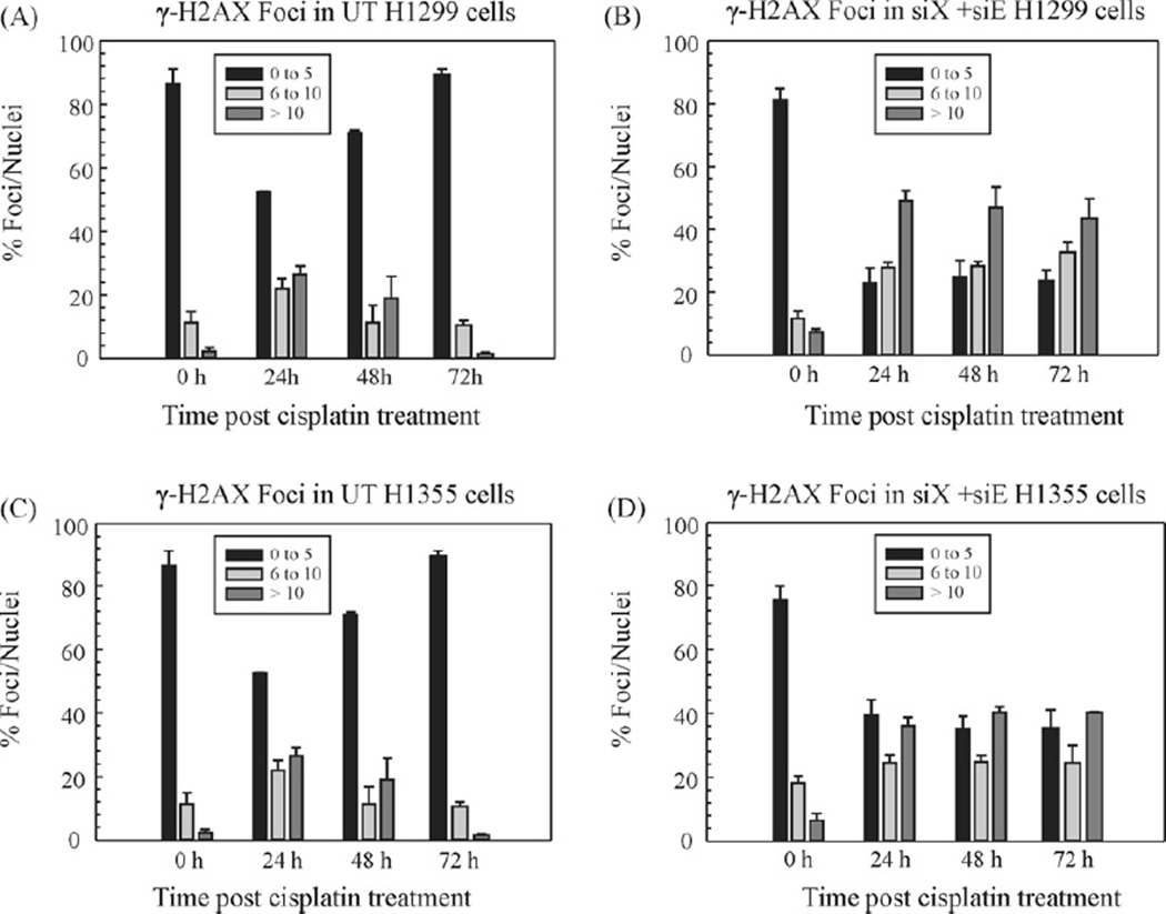
Figure 5.
Repair kinetics of γ-H2AX foci post-cisplatin treatment. H1299 (A and B) and H1355 (C and D) cell lines and quantitation of γ-H2AX focus formation at various time points post-cisplatin treatment in untransfected (A and C) and XPF + ERCC1 (B and D) double knockdown cells, respectively. The cells were seeded onto glass coverslips at 25% confluency. The next day they were treated with cisplatin for 2 h and then fresh complete medium was added. The cells were fixed and immunostained for γ-H2AX at the indicated time points post-cisplatin treatment. For each data point, foci were counted in 250 cells per condition in each cell line. The foci have been categorized as having 0–5, 6–10, or >10 foci per nucleus. The results are expressed as % γ-H2AX foci per nuclei. The data was collected from two individual experiments.

