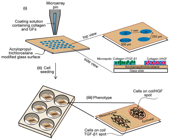
Fig 1. Outline of microarray fabrication.
Collagen I was mixed with HGF or TGF-β1 before being patterned on a glass slide coated with acrylopropyl trichlorosilane. Each spot is 150 μm, 300 μm or 500 μm in diameter and spacing between spots is 1.0 mm (center-to-center) (i). Hepatocytes were seeded onto the glass slides to form a monolayer of cells atop the collagen I spots containing two different growth factors (ii). Schematic diagram showing the morphology of rat primary hepatocytes cultured on HGF and TGF-β1 spots (iii). Abbreviation: GFs, growth factors; col, collagen type I; HGF, hepatocyte growth factor; TGF-β1, transforming growth factor-β1.

