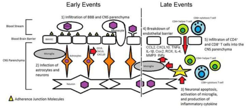
Figure 6.
Infection of CNS by flaviviruses. (1) Infection of the CNS occurs either through the adherence of the virus to molecules present on the surface of brain microvascular endothelial cells (BMVEC's) or infiltration of infected monocytes across the BBB. Viral infiltration then leads to infection of the BBB and CNS cell populations. (2) Infection of human astrocytes leads to chemokine production facilitating further recruitment of monocytes and macrophages. (3) Neurons infected by flaviviruses undergo apoptosis and activates the resident microglia population which produces an inflammatory response. Production of inflammatory cytokines (e.g. TNF-α, IL1β, INF-γ and IL-4), chemokines (e.g. CCL2, CCL5, CXCL9, CXCL10), inflammatory enzymes (COX2) and matrix-metalloproteinases (MMPs) leads to degradation of the endothelial barrier and the release of inflammatory factors (5) recruiting CD4+ and CD8+ T lymphocytes into the CNS parenchyma. Infiltration of CD4+/CD8+ T lymphocytes leads to further inflammation and eventually CNS damage.

