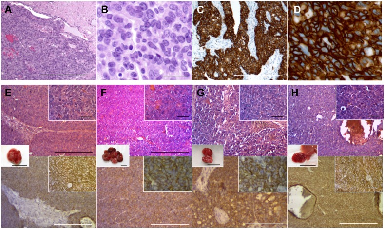
Fig 3. Histology of the patient and mouse-grown tumors and metastasis.
(A) H&E-stained section of the original patient tumor. (B) High magnification image of (A). (C) Immunostained section of the original patient tumor using anti-HER-2 antibody. (D) High-magnification image of (C). (E-H) H&E-stained and immunostained sections of mouse-grown tumors. Upper panels are H&E-stained sections and lower panels are immunostained sections using an anti-HER-2 antibody. Right upper insets are high magnification images (scale bars: 25 μm) and the insets between upper and lower panels are images of the tumors (scale bars: 10 mm). All mouse-grown tumors, including the subcutaneous tumors (E), primary orthotopic tumor (F), peritoneal-disseminated metastasis (G) and liver metastasis (H) had histological structures similar to the original patient tumor and were stained by an anti-HER-2 antibody. Scale bars: 200 μm (A, C, E—H) and 25 μm (B and D).

