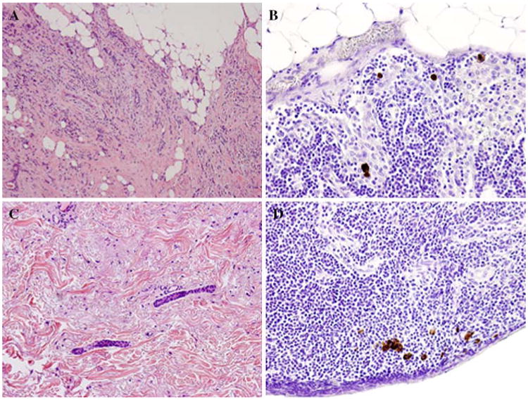
Fig. 1.

Lobular histology and lymphovascular invasion (LVI) in the primary tumor are associated with the detection of isolated tumor cells (ITC) in patients with early-stage breast cancer. (A) Hematoxylin and eosin (H&E) staining of a tumor from a patient with early-stage breast cancer demonstrating lobular histology. (B) Immunohistochemical (IHC) staining for cytokeratin of a sentinel lymph node (SLN) from the same patient demonstrates scattered foci of ITC staining brown. (C) H&E staining of a second patient's primary tumor demonstrating LVI. (D) IHC performed on the SLN from the second patient showing ITC.
