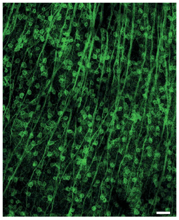
Fig 3.

Channelrhodopsin-expressing cells in neural tissue. The tissue is mouse retina; the ChR2-expressing cells are the retinal ganglion cells; scale bar = 50 μm (image adapted from [2]). Note the high-density of the ChR2-expressing cells; approximately 1/3 of the cells in the population express the protein. This illustrates the need for a device capable of high density, multisite stimulation.
