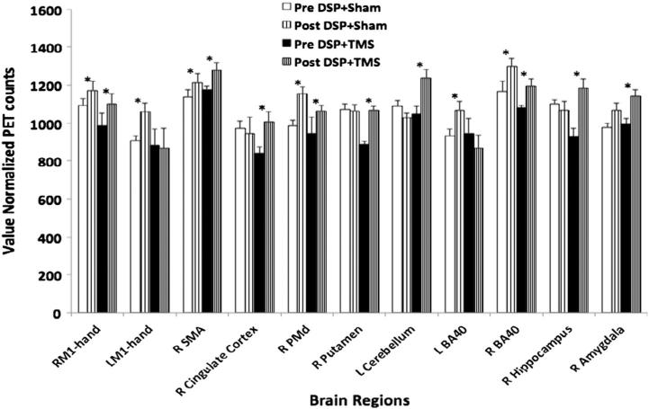
Fig. 7.
Right: resting state CBF amplitude changes following 4 weeks of DSP + sham and DSP + TMS. TMS was applied to right M1hand area. Significant increases in rsCBF were noted in right M1hand, supplementary motor area (SMA), premotor cortex (PMd), and inferior parietal lobule (BA 40) in both groups after 4 weeks of DSP. The DSP + sham group demonstrated significant increases in the left M1hand, and left BA 40 following training. Additional increases in rsCBF were noted in the right sided cingulate cortex, putamen, hippocampus, amygdala, as well as cerebellum in the DSP + TMS group. * = p < 0.05.

