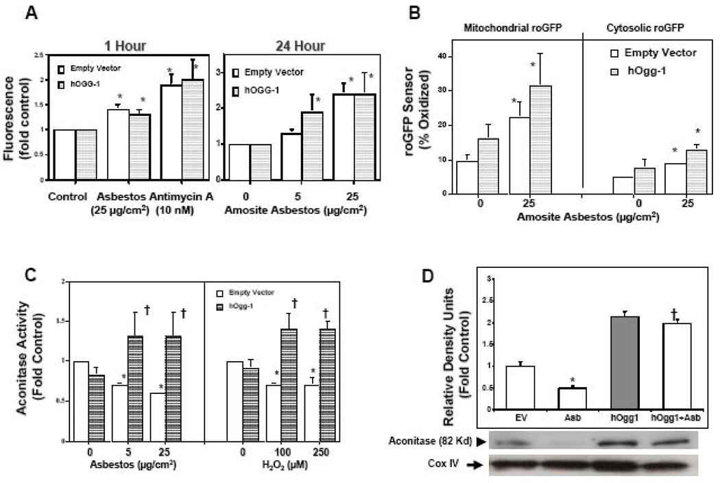
Figure 3. Mitochondria-targeted hOggl does not alter oxidant-induced ROS production but does prevent reductions in mitochondrial aconitase.
(A) Mt-hOggl overexpression does not alter amosite asbestos- or Antimycin A-induced ROS production as assessed by a dichlorofluorescein (DCF) assay. (B) Mt-hOggl overexpression does not block asbestos-induced ROS production as assessed by a roGFP sensor targeted to the mitochondria or cytosol. (C) As expected, mitochondrial aconitase activity is reduced by oxidative stress (24 h exposure to amosite asbestos or H2O2). Notably, mt-hOggl overexpression completely preserves mitochondrial aconitase activity in the setting of oxidative stress. (D) Amosite asbestos decreases mitochondrial aconitase protein expression and mt-hOggl overexpression blocks this. The levels of mitochondrial aconitase expression from three experiments are shown in a densitometric analysis. Expression of cytochrome oxidase IV (COX IV) was used to confirm the presence of mitochondrial protein and comparable loading. * p < .05 v. empty vector controls not exposed to H2O2, Antimycin A, or asbestos; † p < .05 v. empty vector cells exposed to the same dose of H2O2 or asbestos.

