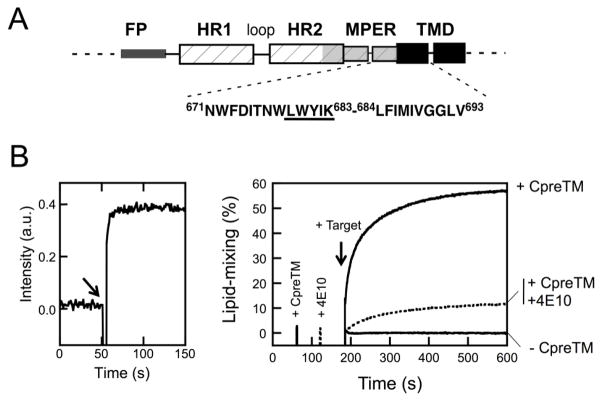
Figure 1. Fusion-committed state generated in vesicles by CpreTM.
A) Designation of the HIV-1 gp41 MPER-TMD connecting sequence. The schematic diagram designates gp41 regions: FP, fusion peptide; HR1 and HR2, amino- and carboxy-terminal helical regions, respectively; MPER, membrane-proximal external region; TMD transmembrane domain. The CpreTM peptide sequence covers Env residues 671–693 (below). B) Left: kinetics of CpreTM incorporation into vesicles monitored by measuring energy transfer from tryptophans to membrane-residing d-DHPE. The peptide was added to a vesicle suspension (100 μM lipid) at the time indicated by the arrow (t = 50 s). The peptide-to-lipid ratio was 1:150 in this assay. Right: Lipid-mixing assay based on the use of fusion-committed vesicles. Unlabeled POPC:Chol (1:1) vesicles (90 μM lipid) were primed at t = 60 s with CpreTM under conditions allowing fast incorporation of the peptide (indicated by the 1st intensity spark). Further co-incubation with the fluorescently-labeled target vesicles devoid of peptide (10 μM lipid) resulted in the mixing of the constituent lipids of both types of vesicles, monitored as the dilution of the probes into the whole vesicle population (arrow at t = 180 s). The final CpreTM-to-lipid ratio was 1:300. Control blank vesicles without CpreTM underwent no lipid-mixing upon target vesicle addition (−CpreTM trace). The dotted trace follows lipid-mixing of CpreTM-primed vesicles that were treated with MAb4E10 (10 μg mL−1) prior to target vesicle addition (2nd intensity spark at t = 120 s). Lipid-mixing was strongly attenuated in this case.

