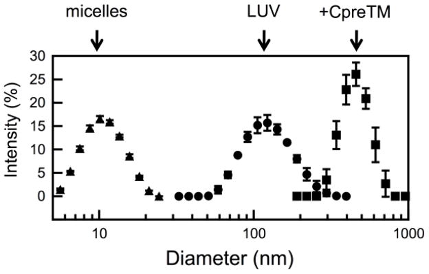
Figure 3. Size distribution of the vesicles determined by light scattering.

POPC:Chol (1:1) vesicles were measured before (LUV, circles) and after 15 min incubation with CpreTM (peptide-to-lipid ratio of 1:100) (+CpreTM, squares). Vesicles treated with Triton X-100 (10 % v/v) are shown as a reference for the size of micelles (micelles, triangles).
