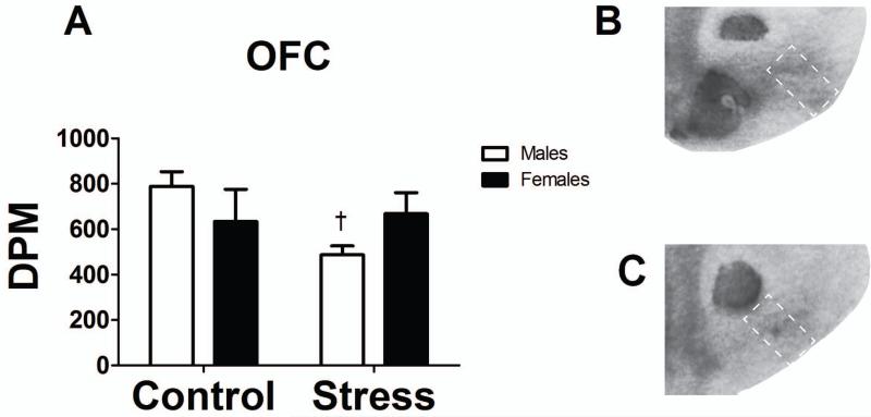
Fig. 4.
(A) Bar graph representing the disintegrations per minute (DPM) for MOR binding in the lOFC in socially defeated or control mice. White bars represent males and black bars represent females (n = 4-5 mice). (B) A representative image of the lOFC in a control male depicting MOR binding following the autoradiography assay. (C) A representative image of the lOFC in a defeated male depicting MOR binding following the autoradiography assay. White dotted boxes represent the region of interest (lOFC).
†p < 0.05 within sex comparison

