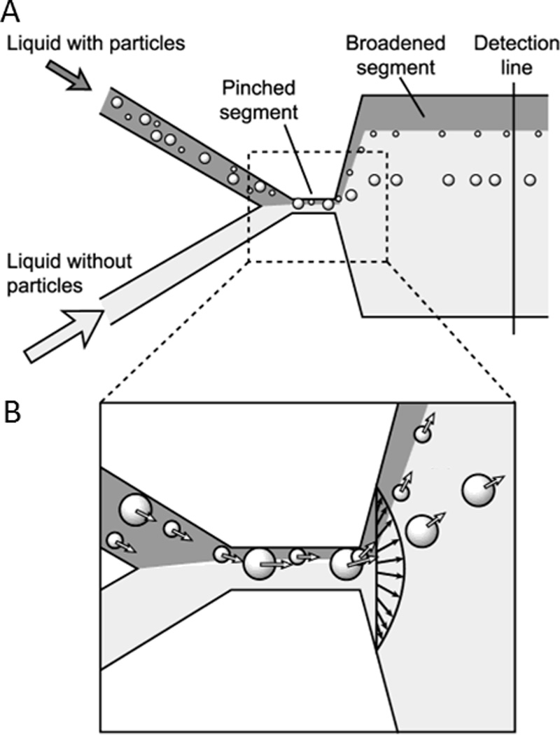
Fig. 11.

Cell sorting by pinched flow fractionation. A) In the pinched segment, cells are first pushed against the wall, and then separated by size upon broadening of the microfluidic channel. B) Cells are aligned in the pinched segment of the channel and follow separate streamlines for sorting by size after exiting the pinched segment. Reprinted with permission from Yamada et al167 Copyright 2004 American Chemical Society.
