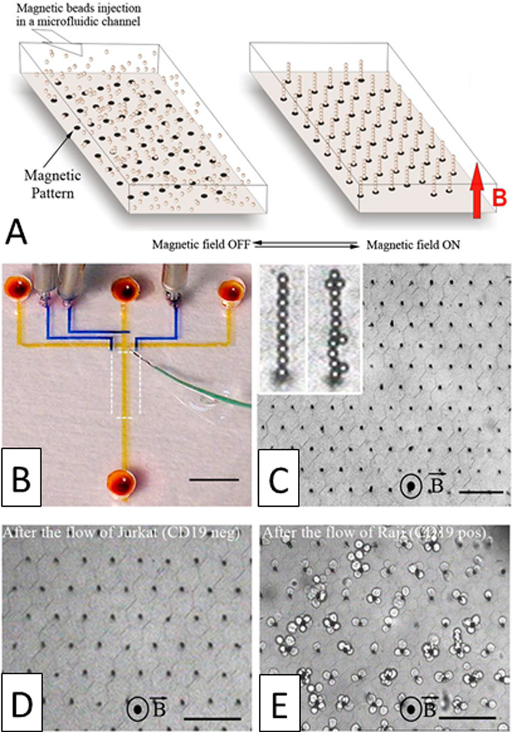
Fig. 18.

Magnetic self-assembly of biofunctional magnetic beads for isolating rare cells. A) Schematic of a hexagonal array of magnetic ink (left) can guide the self-assembly of magnetic beads conjugated with anti-CD19 mAb in the presence of a vertical magnetic field (right). B) Photograph of the microfluidic device. Optical micrographs of the columns after C) the assembly of magnetic beads, D) the passage of 1,000 Jurkat cells (CD19 negative), and E) the passage of 400 Raji cells (CD19 positive) (scale bar: 80 µm). Reprinted with permission from Saliba et al224 Copyright 2010 Proceedings of the National Academy of Sciences of the USA.
