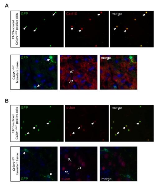
Fig. 2. FACS-enriched transcripts are not expressed in GFP+ microglia in situ.
Immunofluorescence analysis of FACS-sorted Cx3cr1+/GFP microglia and Cx3cr1+/GFP tissue cryosections using Cxcl10 (A) and c-Jun (B) antibodies shows that FACS-sorted GFP-positive microglia express these proteins, whereas GFP-positive microglia in Cx3cr1+/GFP tissue sections do not express these proteins. The nuclei were counterstained with DAPI (blue). Representative images are shown.

