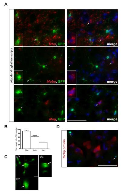
Fig. 3. Fluorescent in situ hybridization confirms the localization of oligodendrocyte-specific transcripts in Cx3cr1+/GFP microglia.
(A) RNA FISH reveals oligodendrocyte-specific (Mbp, Mobp, Mag) mRNA punctae (red) in 34-91% (B) of GFP+ microglia. Representative images are shown with insets of GFP-positive microglia as well as GFP-negative cells containing mRNA molecules. Scale bar, 50μm. (C) A representative high resolution confocal z-stack projection demonstrates Mag mRNA puncta in GFP-positive microglia. Arrow points to the co-localization of mRNA and GFP fluorescence (yellow color). x/y, x/z and y/z projections are shown to confirm the intracellular localization of mRNA within a microglial cell body. Scale bar, 5μm. (D) Immunofluorescence analysis of paraformaldehyde-fixed tissue cryosections using Mobp antibodies (red) and endogenous GFP (green) shows that the Mobp mRNA present in Cx3cr1+/GFP microglia is not translated (arrows). The nuclei were counterstained with DAPI (blue). Representative images are shown. Scale bar, 50μm.

