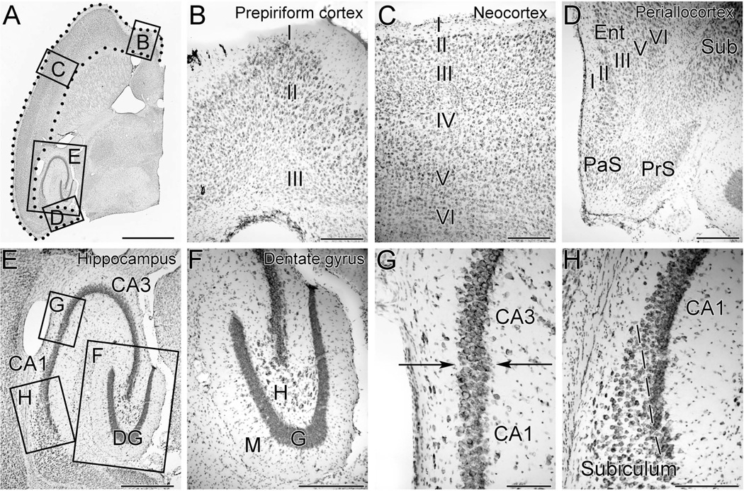
Figure 1. Photomicrographs of a Nissl-stained 40-µm-thick horizontal section of mouse forebrain at a mid-temporal plane demonstrating the boundaries of brain regions examined in this study.
A. The cerebral cortex included all cortical tissue extending from the interhemispheric fissure anteriorly to the subiculum posteriorly and is demarcated by the region within the dotted line.
B. Higher magnification view of the box B in panel A demonstrates the prepiriform cortex, a three-layered cortex, located anteriorly within the cerebral cortex.
C. Higher magnification view of the box C in panel A demonstrates the neocortex, which consists of six distinct cellular layers and constitutes the bulk of the mouse cerebral cortex.
D. Higher magnification view of the box D in panel A demonstrates the periallocortex, a five-layered cortical structure that includes the entorhinal cortex (Ent). The parasubiculum (PaS) and presubiculum (PrS) were also included in the measurements of cerebral cortex, while the more anterior subiculum (Sub) was not.
E. Higher magnification view of the box E in panel A demonstrates the subregions of the hippocampal formation, including the dentate gyrus (DG), CA3 pyramidal cells and CA1 pyramidal cells.
F. Higher magnification of box F in panel E demonstrates that the granule cells of the dentate gyrus (G) can be easily distinguished from cells of the molecular layer (M), and hilus (H).
G. Higher magnification of box G in panel E demonstrates the border between CA3 and CA1 (arrows). CA3 pyramidal cells have larger cell bodies and a lower packing density than the pyramidal cells of CA1.
H. Higher magnification of box H in panel E demonstrates the border (dashed line) between CA1 and the subiculum. The CA1 pyramidal cells abut one another and have a higher packing density than do the cells of the subiculum.
Scale bars represent 2 mm in A, 200 um in B-D, 500um in E, 300 um in F, 100 um in G, and 200 um in H.

