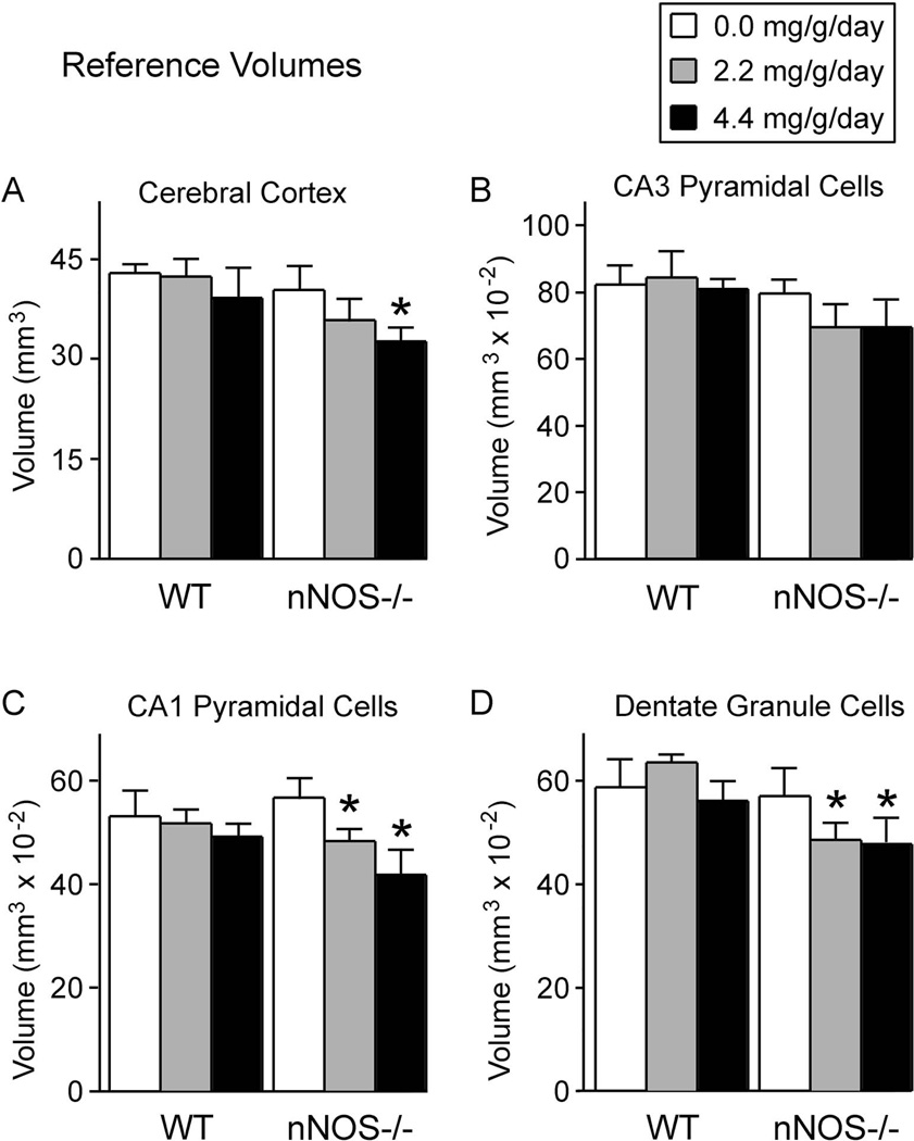
Figure 6. Alcohol reduced reference volumes, but only in nNOS−/− mice.
Shown here are the volumes in which neurons were distributed (the reference volumes), determined by the Cavalieri method, for neurons of the cerebral cortex (A), CA3 hippocampal subregion (B), CA1 hippocampal subregion (C), and the dentate gyrus (D). In the absence of alcohol, reference volumes in the nNOS−/− mice were equivalent to those of wild type mice. Thus, absence of nNOS gene function alone did not affect the volumes of these forebrain regions. In contrast, in the presence of alcohol, reference volumes were reduced, but only in the nNOS−/− mice. Thus, the enhanced alcohol-induced neuronal losses in the nNOS−/− mice were reflected principally by reductions in reference volumes, rather than by reductions in cellular densities.

