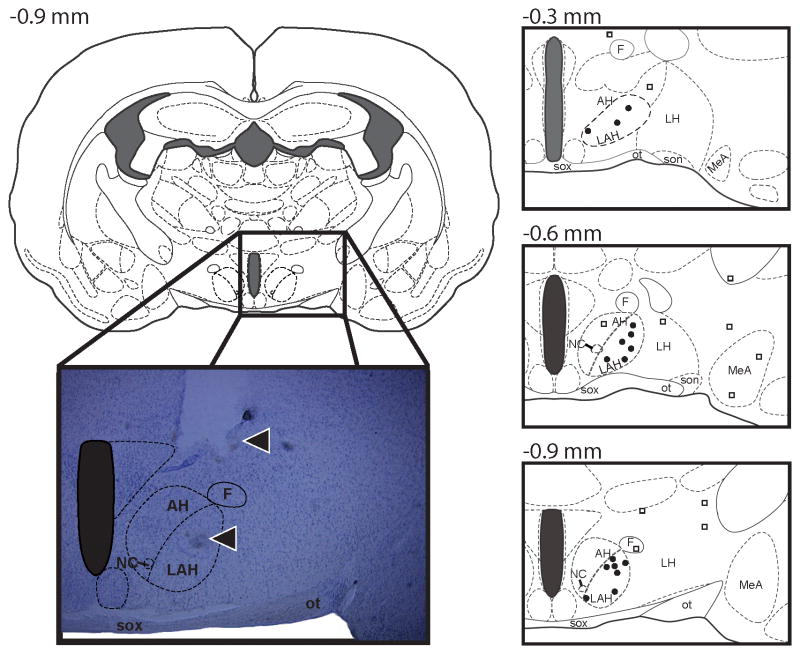
Figure 1.
A series of three atlas schematics at various coronal positions (Morin and Wood, 2001) and a representative photomicrograph of a coronal section of the Syrian hamster brain indicating the sites of central administration of muscimol into the latero-anterior hypothalamus (LAH). The symbol (●) undicates an injection “hit” within the LAH, while the symbol (□) indicates an injection “miss” outside of the LAH target zone. Note in the photomicrograph (from the dorsal to ventral axis) the track of the guide cannula and injection needle (arrows). AH, anterior hypothalamus; F, fornix; LAH, lateral anterior hypothalamus; LH, lateral hypothalamus; MeA, medial amygdala; NC, nucleus circularis; SOX, optic chiasm, ot, optic tract.

