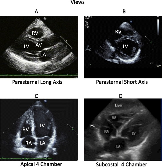
Figure 2.

Standard four views of goal-directed echocardiography. (A) Parasternal long-axis view. Horizontal view of the heart, including the ascending aorta, the aortic valve (AV), the right ventricle (RV), the left ventricle (LV), the left atrium (LA), frequently excluding the apex, and the pericardium. (B) Parasternal short-axis view. A transverse view of the mid-LV at the level of the papillary muscles, including the RV, and the pericardium. (C) Apical four-chamber view, including the LV, RV at the RV inlet, trabeculated apical, and the infundibulum or smooth myocardial outflow regions, LA, right atrium (RA), mitral valve, tricuspid valve, and pericardium. (D) Subcostal four-chamber view. A more perpendicular orientation of the LV, RV, LA, RA, mitral and tricuspid valves, interatrial septum, pericardium, inferior vena cava (IVC), and liver.
