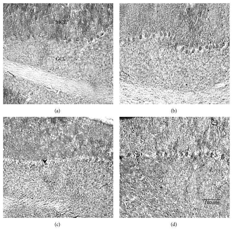
Figure 6.
Photomicrographs of the TrkB stained cerebellums from mutant animals exercised for 7 days (b) or 30 days (d) and their sedentary controls (a) and (c), respectively. Notice that acutely treated runners in (b) have significantly darker stained Purkinje cells than sedentary controls in (a). MCL: molecular cell layer; GCL: granule cell layer. Scale bar in the lower right panel refers to all photos.

