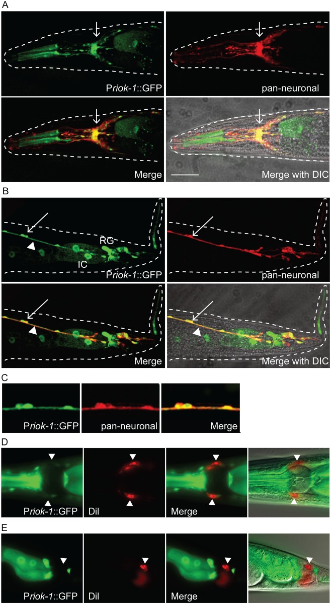
Fig 3. Priok-1_2SKN-1 drives GFP expression in a small subset of neurons.
(A–C) Single plane confocal images from adult worms. (A) GFP was expressed in a subset of head neurons and is highly expressed in the nerve ring displaying strong co-localisation with the pan-neuronal marker (arrow). (B) Co-localisation of GFP with pan-neuronal dsRED. D-type neurons (arrow) and longitudinal nerves (arrow head). Intestinal cells (IC) and the rectal gland (RG) can also be observed. (C) Zoomed in image of the D-type neurons displaying co-localisation. Scale bar = 25μm. (D–E) Priok-1_2SKN-1 GFP expression does not co-localise with the lipophilic dye Dil, in head neurons (D) or tail neurons (E). Scale bar = 15 μm.

