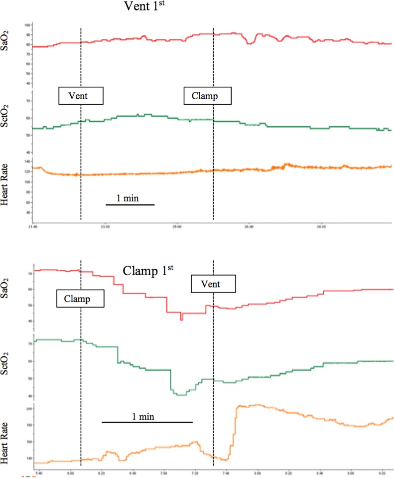
Fig 2. Effect of the timing of ventilation onset relative to umbilical cord clamping.
Representative traces obtained from a lamb in which ventilation was initiated prior to umbilical cord clamping (Vent 1st), and a lamb in which umbilical cord clamping was conducted prior to the initiation of ventilation (clamp 1st). Dashed line indicates when an intervention occurred as labeled on the graphs. Note the difference in time scale. SpO2—arterial oxygen saturation, SctO2—cerebral oxygenation.

