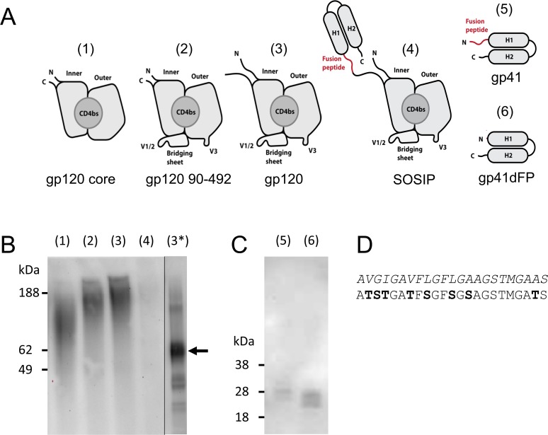
Fig 1. Expression analysis of different gp140 fragments.
(A) Schematic representations of expressed HIV-1 gp140 fragments, including gp120 inner and outer domain (inner and outer), CD4 binding site (CD4bs), V1/2 loop region (V1/2), the V3 loop (V3) and helical regions 1 and 2 of gp41 (H1, H2). (B) Western blot analysis of (1) yeast-secreted YU2 gp120 core, (2) gp120 residues 90–492, (3) gp120, (4) JR-FL SOSIP gp140, (3*) PNGaseF-treated YU2 gp120 with marked main band. Lane (3*) is non-adjacent but originates from the same blot. (C) Western blot analysis of (5) yeast-secreted JR-FL gp41 and (6) gp41 lacking the fusion peptide. (D) Amino acid sequences of the original and the modified fusion peptide, with modified residues in bold text.

